Story highlights
Surgeons in Miami use 3-D-printed model of a girl's heart to plan her complicated surgery
Simulated organs are being used to prepare and practice for many operations
Eventually, 3-D printers could produce organs that can be transplanted into patients
Mia Gonzalez spent the first 3? years of her life missing out. She had to skip day care and dance classes because she constantly had colds and pneumonia. When Mia could go out and play, she was easily winded and took multiple asthma medications to try to help her breathing.
After about 10 hospital stays, doctors realized that Mia had a malformation in her aorta, the vessel that pumps blood from the heart. The 4-year-old would need an operation to close off the part of her aorta that was putting pressure on her windpipe and making it hard to breathe, swallow and get rid of phlegm when she got a cold.
“We freaked out to go from thinking she had asthma to being told she needed to have open heart surgery,” said Katherine Gonzalez, Mia’s mom.
But Mia’s malformation was complicated. The surgeons at Nicklaus Children’s Hospital in Miami, where Mia was treated, might have been apprehensive about the procedure were it not for a new technology: the 3-D printer.
No longer ‘inoperable’
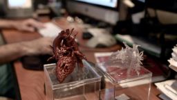
Earlier this year, the hospital got a 3-D printer that makes exact replicas of organs that doctors can use to plan surgery, and even do practice operations. The printer uses images from patients’ MRI or CT scan images as a template and lays down layers of rubber or plastic.
Dr. Redmond Burke, director of pediatric cardiovascular surgery at Nicklaus Children’s Hospital, meditated on the model of Mia’s heart for a couple of weeks. He showed it to colleagues for their input and even carried it around in his gym bag for quick reference.
Burke finally had the “Aha!” insight. Instead of making an incision on the left side for this type of heart defect, called double aortic arch, he should cut into Mia’s chest from the right.

“Without the model, I would have been less certain about (operating on Mia) and that would have led me naturally to make a larger incision that could possibly cause more pain and a longer recovery time,” Burke said.
Using the model, “there was no doubt, and surgeons hate doubt,” he added.
He attributes the model with saving the team and patient about two hours in the operating room because he was able to have a clearer plan to do the surgery.
Burke and his colleagues have created models for about 25 young patients with congenital heart defects. In the past, they might not even have operated on some of these patients because the surgeries seemed too tricky, but “it’s very unlikely I will ever call someone inoperable (now) without holding a model in my hand,” Burke said.
About 75 hospitals in the United States, and around 200 worldwide, have a printer such as the one used to make a model of Mia’s heart, said Scott Rader, general manager of Stratasys, which sells the printers.
Although 3-D printers have been used clinically for the last 20 or 25 years to make prototypes for surgical tools and other uses, they only started churning out simulated organs in the last few years, Rader said.
Doctors have used the simulated organs to prepare for all kinds of tricky operations, such as surgery to remove a brain tumor or to correct a severe cleft palate, Rader said. Doctors can operate on them with regular surgical tools “again and again and again until you think of the optimal way to do surgery,” Rader said.
‘The biggest secret that nobody knows about’
The models can also help train medical students and explain complicated procedures to patients such as Mia and her family.
Eventually, the printers could be used to produce actual organs that can be transplanted into patients; just replace the rubber and plastic printer “ink” with human cells.
“We are going to see massive advances in the next five to 10 years of how to take the technology to this point,” Rader said.

Simulated organs are “a disruptive technology which radically changes how we talk to patients, how we prepare for an operation, how we do the operation and how we teach,” said Dr. Daniel B. Jones, professor of surgery at Harvard Medical School and co-director of the Simulation and Skills Center at Beth Israel Deaconess Medical Center in Boston.
However, the technology is not widely in use in hospitals or medical schools yet.
“This is like the biggest secret that nobody knows about,” said Jones, who has done research on surgeons’ impressions of using simulated organs. Even those who do know about the technology might feel uncomfortable showing patients a model of their tumor, for example, and discussing it with them in depth.
Hospitals are starting to invest in the technology, which is more realistic now that prices have come down, Jones said.
The printer and software usually cost in the range of $100,000, which is less than a CT scan or MRI setup, Rader said. He predicts that interest in the technology will continue to grow as research shows how using simulated organs leads to better surgical outcomes and shorter operating times.
The models have also been invaluable in helping patients and their families feel confident that they are doing the right thing, said Burke, who operated on Mia.
Seeing the model “made it seem like a simple surgery, it put you at ease,” said Gonzalez, Mia’s mom. “(Mia) was used to hospitals and IVs, but to explain that you’re going to have surgery and be in pain, that is all different,” and the model helped put it in perspective, she said.
Four months later, the surgery seems like ancient history to Mia. She has forgotten all about her surgical scar, her mother said, and had little pain. Although she has had some minor colds, none has landed her in the hospital. A month later, she was even able to participate in her dance recital.


